
Utilization of CBCT in pediatric traumatology: the first professional presentation in Slovakia

The use of CBCT devices in traumatology was officially presented to the Slovak medical community for the first time at the 11th international congress “Trauma in Childhood” in late June 2026 at the X-BIONIC Sphere Šamorín.
The professional lecture, delivered by a team of specialists from Medirad (V. Neuschl and A. Uhrinová), received a positive response from the attending pediatric traumatologists and surgeons. Practical demonstrations and direct comparison of image details with minimal radiation exposure confirmed that Slovak pediatric traumatology now holds a new and justified diagnostic tool.
What is CBCT and how does it differ from conventional CT?
If you have ever had a detailed 3D scan of your teeth performed by an orthodontist, you have likely encountered CBCT. For many years, this technology (cone-beam computed tomography) was used almost exclusively in dentistry. However, the latest generation of devices has pushed boundaries and can now rapidly scan any part of the human body, from elbows and wrists to ankles and the spine.
The main difference from conventional “large” CT (so-called MSCT) lies in how radiation passes through the body:
Conventional CT emits a narrow, fan-shaped beam that spirals around the patient’s body, scanning layer by layer.
CBCT uses a cone-shaped beam. The device performs just a single rotation around the injured area, instantly capturing the entire three-dimensional volume of that region.
Comparison of MSCT/CBCT
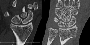
Why CBCT is the ideal choice for children
If a pediatric patient requires a CT scan of the musculoskeletal system, CBCT is definitely the superior choice. Children’s bodies are extremely sensitive to ionizing radiation. Cells in the growth and division phases react much more sensitively to radiation than adults do, and the risk of future health complications imposes strict limits as the child’s age decreases. Therefore, doctors always apply the ALARA principle (As Low As Reasonably Achievable), using the lowest possible radiation dose for diagnosis.
CBCT offers three key advantages compared to conventional CT
Radiation exposure under strict control
Switching from conventional CT to modern CBCT means a dramatic reduction in radiation dose for the pediatric patient. While conventional CT can emit a significantly higher dose than a standard X-ray when imaging a joint, CBCT values remain only slightly above the level of a standard RTG image. Meanwhile, the doctor receives a complete 3D model instead of a flat 2D image.
Extreme detail to reveal “hidden” fractures
Children’s bones are unique, they contain growth cartilage, and injuries often result in so-called occult (hidden) fractures or avulsions (tearing off of small bone fragments). CBCT offers enormous spatial resolution (in the order of hundredths of a millimeter). This allows orthopedists and traumatologists to see even the finest micro-cracks in the bone structure that a standard X-ray would miss.
No image artifacts caused by metal
If a child already has surgical screws, wires, or plates in the bone from previous surgeries, conventional CT creates artifacts around them (the image is “blurred” and distorted near the metal). CBCT technology can suppress these metal artifacts to a minimum. This allows the doctor to precisely check whether the bone is healing correctly under the implant.
Practical application in pediatric injuries
In pediatric traumatology, CBCT should be indicated for complex limb injuries where it is necessary to fully understand the anatomy of the injury before planned surgery or during post-operative checks. Typical examples include:
Elbow and wrist injuries:
Very common in children after falls from bicycles, scooters, or playground equipment. Accurate assessment of fractures in the elbow joint area is crucial for preserving full hand mobility in the future.
Knee and ankle injuries in young athletes:
Sudden rotations and sprains can damage growth plates, which may affect the future growth of the limb without timely diagnosis.
Conclusion
The first professional presentation of CBCT use in pediatric traumatology in Slovakia confirmed the growing interest in this technology. High spatial resolution combined with low radiation exposure represents a significant contribution to the diagnosis of pediatric injuries and expands the possibilities of imaging examinations in daily clinical practice.
The Medirad team will continue to support professional discussion on the use of CBCT and develop its availability for referring doctors and patients. https://www.medirad.sk/vysetrenia/#cat-cbct-3d-skelet