
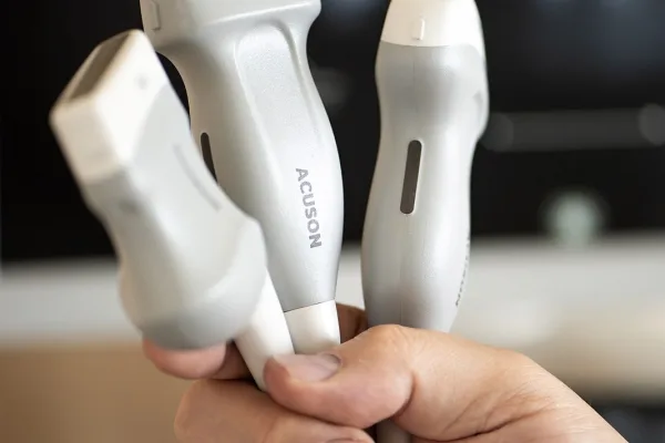
Ultrasound imaging visualizes the inside of the body using sound waves. A probe placed on the skin emits a wave with a frequency above the threshold of human hearing into the tissue and captures what returns. The device constructs an image from this data, which the physician evaluates in real time, i.e., directly during the examination.
Ultrasound, sonography, and the abbreviation USG refer to the same examination. It non-invasively visualizes internal organs, soft tissues, blood vessels, and other body structures.
Ultrasound is safe
Ultrasound does not use radiation. This is the difference from X-ray and CT, which send radiation into the body with enough energy to eject electrons from atoms. Sound waves do not have such energy, so there is no dose in sonography that needs to be tracked or accumulated over time. Therefore, USG examinations can often be repeated without any risks.
This leads to several practical implications:
- The examination can be repeated after a week, a month, or even six months, as many times as the course of the disease requires. This is crucial for monitoring healing or the progression of findings.
- It is performed at any age, from newborns to seniors.
- During pregnancy, it monitors fetal development, precisely where caution regarding radiation is highest.
- Diagnostic sonography does not require a needle or contrast agent. The examination is performed through the skin.
How an image is formed from reflected sound
The probe emits short pulses of high-frequency waves into the tissue. At each interface between media with different acoustic properties, part of the wave reflects back to the probe. The device measures how long each reflection took to return and determines the depth at which the interface lies based on this time. An image is formed from a large number of such measurements and is continuously updated.
Why there is no single probe for everything
The frequency of the wave determines two things simultaneously, which are in conflict with each other. The higher the frequency, the finer the detail the probe can resolve, but the shallower the wave penetrates into the tissue. Lower frequencies penetrate deeper at the cost of a coarser image.
Therefore, the physician selects the probe depending on which area is being examined and at what depth below the surface it lies. Probes differ in shape, frequency range, and method of use.
Linear probe for structures just under the skin
It works with higher frequencies, allowing it to resolve even fine details in the shallow layer beneath the body’s surface. At our clinic, we use it to examine:
- the thyroid gland and soft parts of the neck
- joints, muscles, and tendons
- peripheral nerves
- breasts
- vessels, including carotids as well as veins and arteries of the limbs
Convex probe for deeper organs
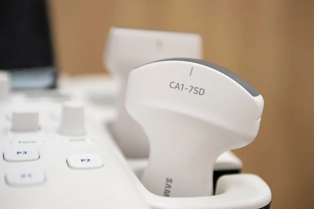
Its curved contact surface provides a wider field of view, and lower frequencies penetrate deeper into the body. Therefore, it is used for examining internal organs. At our clinic, this mainly includes abdominal ultrasound (liver, gallbladder, and kidneys) and examination of the urinary tract and pelvic organs.
Sector probe for examination between ribs
It has a small contact surface, allowing it to reach narrow anatomical spaces where there is little room for larger probes. It images, for example, from between the ribs, which is why we use it for chest ultrasound.
Where we perform sonography at our clinic
We perform sonographic examinations in two sonography offices staffed by physicians specialized in radiology. We have multiple types of probes available, so the physician chooses the one that corresponds to the area being examined.